Preimplantation Genetic Diagnosis
Genetic diseases are considered to be a major problem. Concentrated efforts have been done by scientists to solve the problem. We will mention a few of the steps taken.
Pre-implantation diagnosis by embryo biopsy:
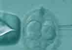
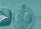
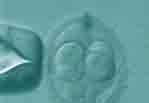
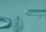
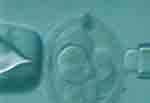
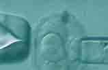
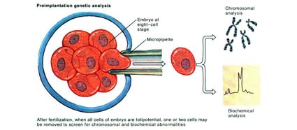
This is done by taking one or more cells from the embryo. Then, scientists try their best to take as many cells as possible to reach a diagnosis or identify a disease. The best time to do this is after the division of the fertilized egg by IVF, until it reaches the stage of 8 cells. That usually occurs on Day 3 after fertilization. The lab technician creates a hole in the embryo wall by using a very thin microscopic needle or chemical substance, or laser. Then, one cell will be taken without causing damage to the embryo by taking the cells that have no effect on the embryo (nonessential cells) in an advanced stage of the embryo's development (the blastocyst stage), and after that, the diagnosis is done in 2 ways:
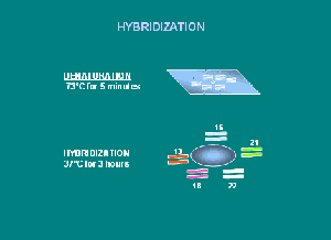
2. FISH (fluorescence in situ hybridization):
In this technique, the normal embryos can be selected to be transferred to the uterus as in the case of IVF. It should be taken in to consideration to select the best time to take the biopsy from the embryo and to avoid occurrence of damage to the embryo. In that way, we can achieve the best results in diagnosis which will lead to a healthy, successful pregnancy.





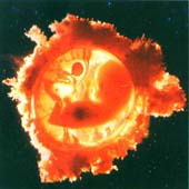
1. Genetic diagnosis of pre-implantation embryo:
This technique is used to diagnose the inherited diseases and has been developed recently, by using a special test called PCR (Polymerase Chain Reaction) and the number of human genetic diseases that can be diagnosed is increased. We hope, in the future, scientists will be able to diagnose all the genetic diseases in that way. This technique is useful for couples that may have a child who is effected with an inherited disease. In that way, only the normal embryos can be transferred to the uterus. This is much better than the occurrence of a pregnancy than the diagnosis of a genetic disease by Amniocentesis (taking sample of the amniotic fluid), or by Chorionic villous sampling.
Gene disorders transmittable to the offspring, which can be analysed by genetic diagnosis after oocyte and embryo biopsy.
|
Achondroplasia |
Central core disease |
|
Agammaglobulinemia |
Gaucer’s disease |
|
Sickle-cell anemia |
Huntington’s disease |
|
Fanconi’s anemia |
Alport’s disease |
|
Spinal/bulbar muscular atrophy |
Tay-Sachs’ disease |
|
Alpha1- antitrypsin deficiency |
MELAS |
|
Long chain hydroxyacyl CoA dehydrogenase deficiency |
X-linked myotubular myopathy |
|
Ornithine transcarbamilase deficiency |
Neurofibromatosis I and II |
|
Deficiency of the mitochondrial trifunctional protein |
Multiple endocrine neoplasia type II |
|
Multiple epiphyseal dysplasia |
Osteogenesis imperfecta I and IV |
|
Myotonic dystrophy |
Familial adenomatous polyposis coli |
|
Becker’s muscular dystrophy |
Rhetinitis pigmentosa |
|
Duchenne’s muscular dystrophy |
Rhesus (Rh D) |
|
Haemofilia A and B |
Tuberous sclerosis |
|
Epidermolysis bullosa |
Crouzon’s syndrome |
|
Exclusion HD |
Di George’s syndrome |
|
FAP-Gardner |
Hunter’s syndrome MPS II |
|
Phenylketonuria |
Lesch-Nyhan’s syndrome |
|
Cistic fibrosis |
Marfan’s syndrome |
|
X-linked hydrocephalus |
Digital oro-facial-syndrome type 1 |
|
Incontinentia pigmenti |
Stickler’s syndrome |
|
Hyperinsulinemic hypoglycemia PHH1 |
Fragile X syndrome |
|
Early onset Alzheimer’s disease |
Wiskott-Aldrich syndrome |
|
Charcot-Marie-Tooth’s disease 1 and 2A |
Thalassemia |
List of some of the genetic diseases that can be diagnosed by PGD
2. FISH (fluorescence in situ hybridization):
Normal embryos can be selected to be transferred to the uterus, as in the case of IVF. It should be taken in to consideration to select the best time to take the biopsy from the embryo, and to avoid the occurrence of damage to the embryo. We can then hope for the best results in diagnosis, leading to a healthy and successful pregnancy.
Dr Najeeb Layyous F.R.C.O.G
Consultant Obstetrician, Gynecologist and Infertility Specialist


 Pregnancy Due Date Calculator
 Chinese Gender Predictor
 Ovulation Calculator
 IVF Due Date Calculator
